Colles Fracture (Distal Radius Fracture)
WHAT IS A COLLES FRACTURE (Distal radius Fracture)
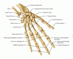
In the medical community, professionals often refer to a distal radius fracture using the term Colles Fracture. Named after Abraham Colles who defined this type of fracture, Cole introduced Colles Fracture in medical literature in 1814. The wrist has two bones in the forearm, the ulna and the radius. With the body in anatomical position, the radius sits lateral to the ulna. Individuals can find the radius on the thumb side of the wrist. The radius rotates around the ulna when the arm twists. Physicians suggest the distal portion of the radius makes up roughly the third of the bone nearest to the hand. The most common of wrist fractures, a distal radius fracture, occurs most often from falling onto an outstretched arm. The second most common way that a distal radius fracture occurs happens from traumatic impact. Frequently the fracture appears non-displaced or slightly displaced and does not require surgical intervention. If the forearm experiences an out of place bone, a medical provider must set the bone back into place before casting. To reset the bone, the patient may need to undergo surgery. However, if the fracture extends into the wrist joint, breaks through the skin, fractures multiple places, severely dislocates, or peripheral damage occurs to the vascular system or the tendons of the wrist, then the patient may require surgical intervention.
WHAT ARE SYMPTOMS OF A COLLES FRACTURE?
Symptoms of a fracture, Colles or otherwise, typically exhibit similar symptoms with one another. With a fractured bone, pain occurs when using the affected limb, in this case the wrist. The pain may worsen while flexing the wrist. With a radius fracture present, the patient may also feel tenderness in the wrist area, especially while palpating the wrist. Swelling, bruising, and possible deformity of the wrist may also occur with a wrist fracture. The pain may feel like a consistent deep ache with additional sharp pain when moving or irritating the fracture. All of these symptoms in conjunction with a traumatic incident to the wrist should warrant a trip to see an orthopedic hand and wrist specialist. The orthopedic hand and wrist specialist evaluates, reduces, and immobilizes the fracture. If necessary, the orthopedic specialist may recommend surgical intervention.
diagnosing and treating a COLLES, DISTAL RADIUS, FRACTURE
To diagnose a Colles fracture, or Distal Radius Fracture, the orthopedic specialists starts with a physical examination. During a physician exam, the doctor goes over patient history with the patient. The doctor also goes over the daily recreational activities that the patient performs in addition to work activities. Next, the orthopedic hand and wrist specialist examines the affected arm and wrist. The doctor may move the wrist and palpate the wrist in different areas to see which areas reproduce pain. Next, the physician orders diagnostic testing. Often, a simple X-Ray can diagnose a Colles fracture, or Distal Radius Fracture. If the orthopedic specialist finds the Colles fracture in the wrong position to heal, the orthopedic hand and wrist specialist may need to reset the fracture to put the fracture back into the correct alignment. Surgeons often reset fractures with the patient asleep under anesthesia due to the painful process. Medical professionals refer to resetting a fracture using the term fracture reduction. Often, fracture reductions occur in Emergency Rooms.
To reset a Colles fracture, or Distal Radius Fracture, an anesthesiologist places the patient under either local or general anesthesia. Often, doctors can perform a fracture reduction using a nerve block and local anesthesia. If the fracture appears more severe, the doctor may opt for general anesthesia. With a patient under general anesthesia, the patient remains asleep for the entirety of the procedure. While asleep, the patient does not feel anything and does not wake up until after the procedure. Once the anesthesiologist places the patient under, the surgeon performs the reduction. To perform a fracture reduction, the orthopedic hand and wrist specialist moves the end of the broken bone the place it back into proper alignment. Orthopedic specialists must perform fracture reductions in order for the bone to heal correctly. If a Colles fracture, or Distal Radius Fracture goes left untreated and does not get reduced, many issues may arise for the patient in the future. The patient may experience great pain along with nerve or artery damage. Following the reduction, the treating physician must hold the fracture in place so the fracture can heal properly. Initially, the treating physician often prescribes a splint to hold the fracture into place while the swelling goes down. Once the swelling goes down, the doctor most likely fits the patient for a cast a week or so later. The patient must wear the case for roughly six to eight weeks. In many cases, the patient has two casts during their casting period. The doctor changes the cast to inspect the healing progress and replace the cast for hygienic reasons. The cast wearing time frame may vary based on the provider and the specific fracture. Following the hard cast, the physician may require the patient to wear a soft cast for 4 more weeks to ensure the wrist remains protected and stabile. Severe cases of a Colles fracture, or Distal Radius Fracture may require distal radius fixation with a volar plate. Using a volar plate to fixate a fracture requires a surgeon to use pins or screws to hold the reduced fracture into place.
Recovering from a Colles fracture, or Distal Radius Fracture
After the casting period, many muscles surrounding the wrist may have atrophied. To protect the bones of the wrist, the muscles surrounding the wrist must remain strong. To strengthen these muscles post casting, the treating physician may require the patient to participate in physical therapy.
To view a list of all insurances that AOA Orthopedic Specialists accept, click HERE. To schedule an appointment online, click HERE.


